Hermia Advanced Bone SPECT Reconstruction
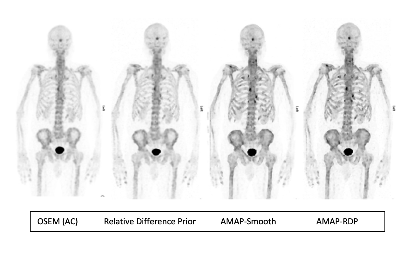
Bone SPECT/CT gains a new level of clarity with Bayesian reconstruction (AMAP) in Hermia. Decrease noise in your clinical bone SPECT/CT routine today and improve lesion quantitation and detectability with no additional hardware requirements. Our easy-to-use software enables precise determination of reconstruction parameters, and we are proud to be able to offer two Bayesian reconstruction algorithms to optimise your bone SPECT/CT images: AMAP-Smooth and AMAP-RDP. Hermia’s advanced reconstruction methods work with current generation scanners from all manufacturers.
Vuohijoki et. al NMC 2023 (1) have shown in their study that anatomically guided Bayesian reconstruction (AMAP) and Kernelized Expectation Maximization (KEM) reconstruction techniques deliver significantly better quantitative accuracy and lesion detectability compared to the commonly used ordered subsets expectation maximization (OSEM) method.
What is it?
- Using prior information about how the final image is expected to look, the reconstruction algorithm is guided towards a clearer, less noisy solution
- Anatomical prior information from the CT can be included to smooth only in uniform tissue areas and avoid smoothing over edges
What is the benefit?
- Higher contrast lesions for bone SPECT/CT
- Less noisy images
- Better quantitative accuracy
Published results
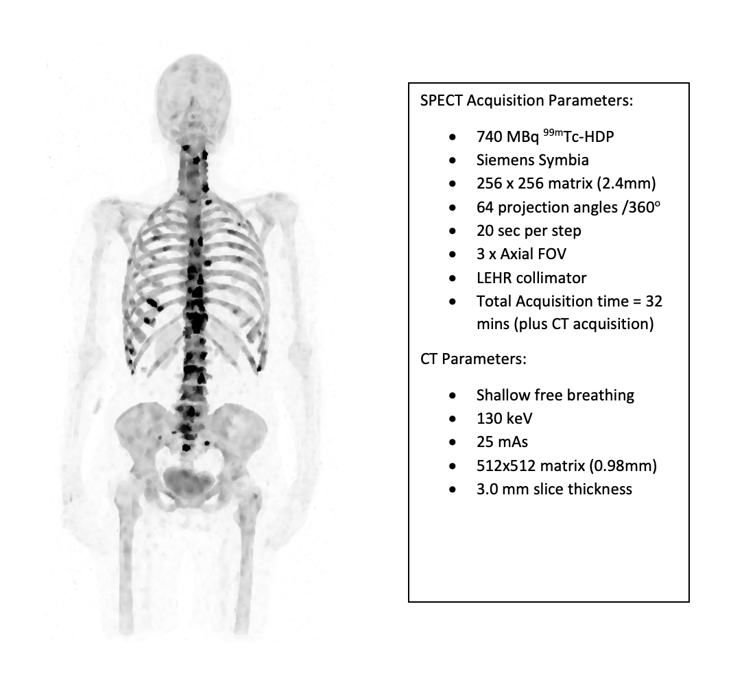
In a recent study (2023)1 undertaken at the Department of Clinical Physiology and Nuclear Medicine at Päijät-Häme Central Hospital in Lahti, Finland, it was demonstrated for the first time that reconstruction using CT information can improve lesion detection in bone SPECT/CT scans.
Artificial lesions with a wide range of lesion-to-background contrasts were added to normal bone SPECT/CT studies. The quantitative accuracy was assessed by the error in lesion standardized uptake values and lesion detectability by the area under the receiver operating characteristic curve generated by a non-prewhitening matched filter.1
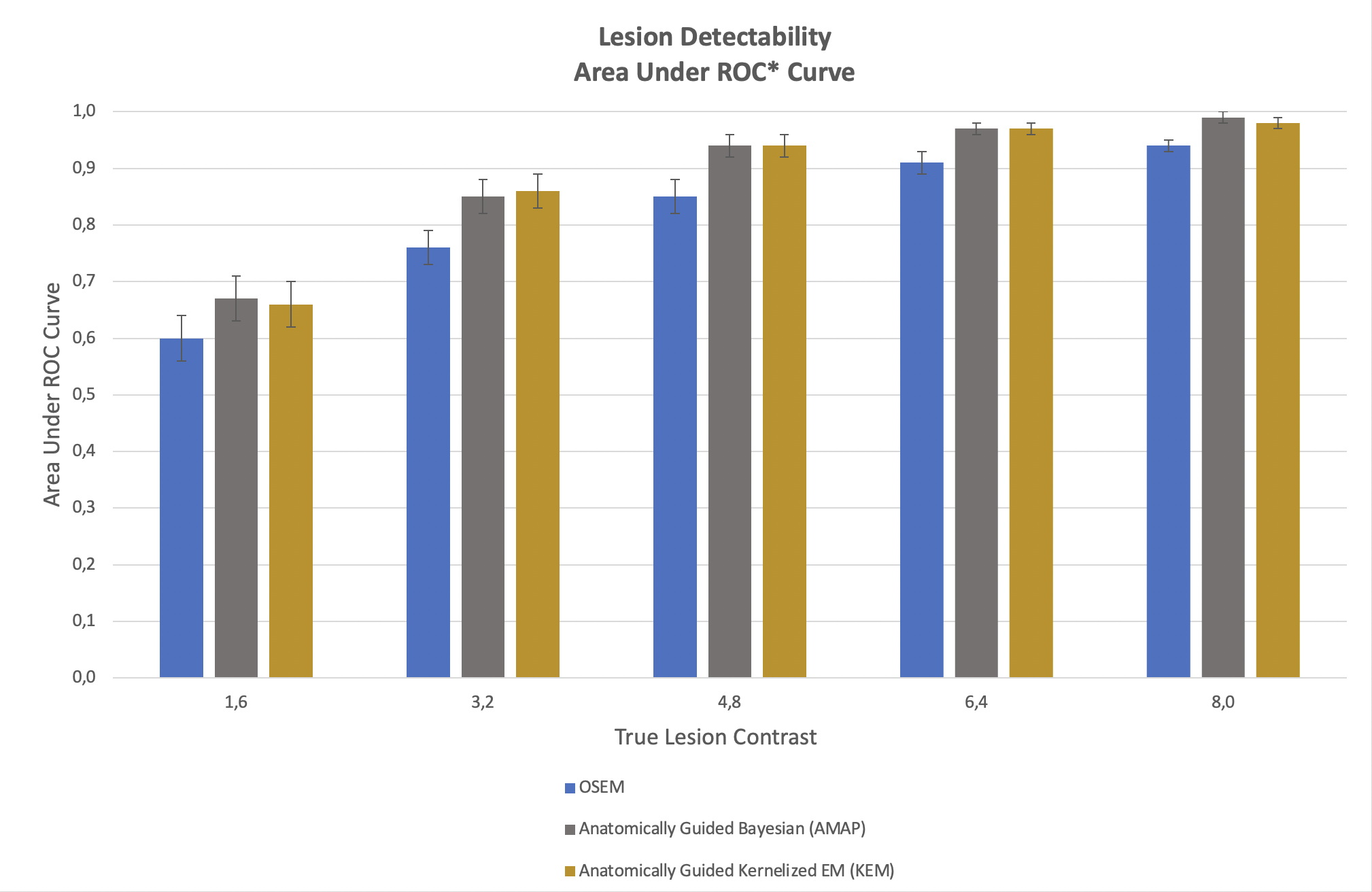
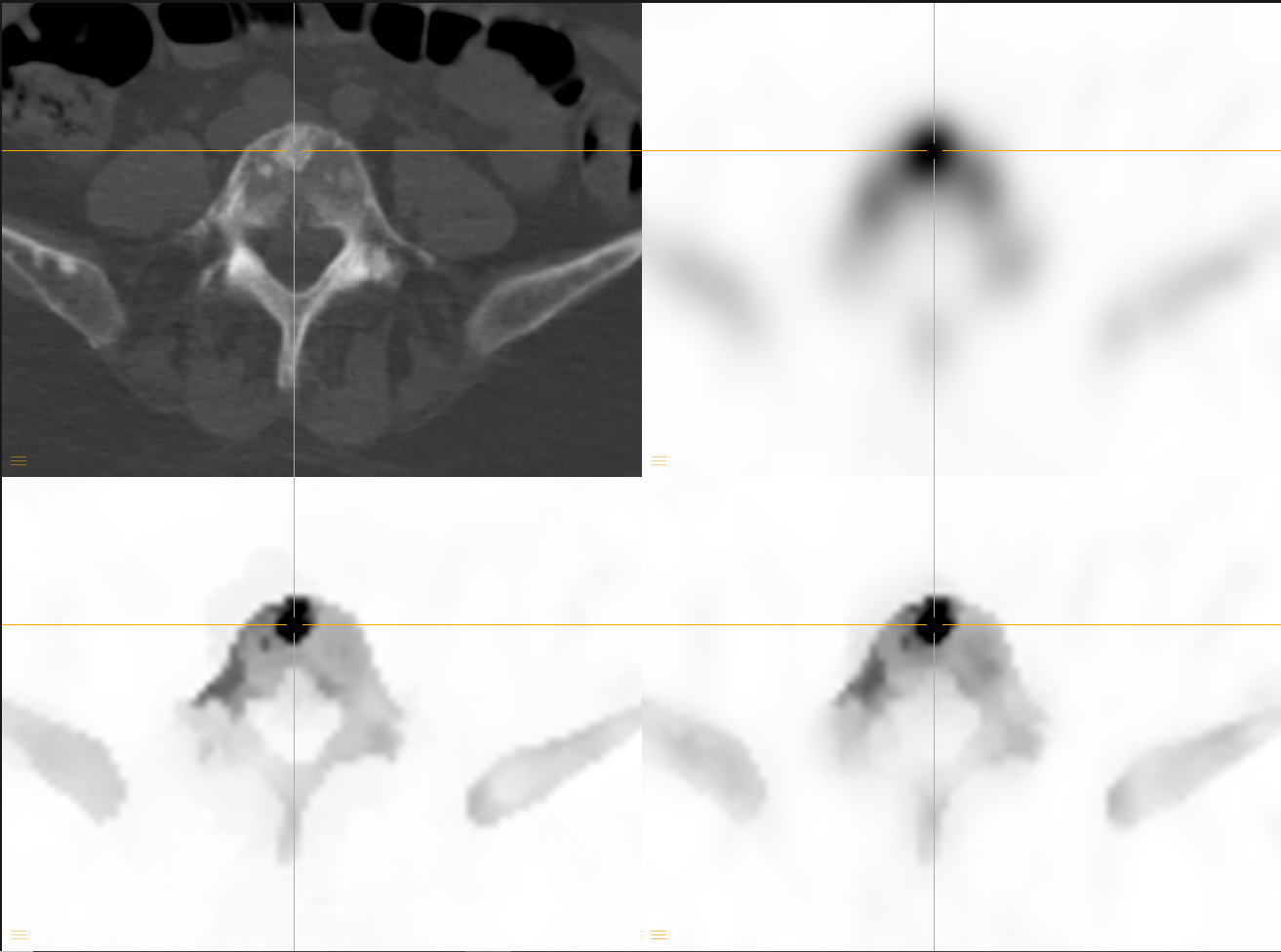
In figure 1, we can see how the two anatomically guided reconstructions methods AMAP and KEM perform better at lesion detection compared to conventional OSEM.
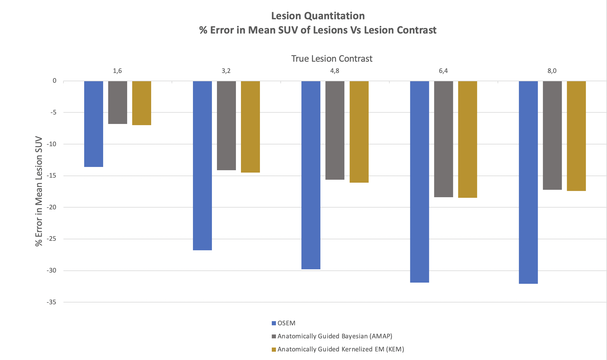
This can also be seen in figure 2. In the study by Vuohijoki et al. (2023) artificial lesions were added to the SPECT data to allow determination of absolute errors in mean standardized uptake value (SUV) per lesion. Figure 3 shows that AMAP and KEM clearly outperform OSEM in terms of mean SUV lesion quantitation accuracy.
In a previous study performed by the team at Vaasa Central Hospital, in Finland2, anatomically guided reconstruction showed to be improving lesion quantitation accuracy in bone SPECT/CT.
Artificial lesions were added to the SPECT data to allow determination of absolute errors in mean standardized uptake value (SUV) for each lesion. The study found the following average relative error in mean SUV for OSEM, RDP, AMAP-S, and AMAP-R to be respectively − 53%, − 35%, −15%, and − 10%, when the CT study had matching lesions, see graph below.
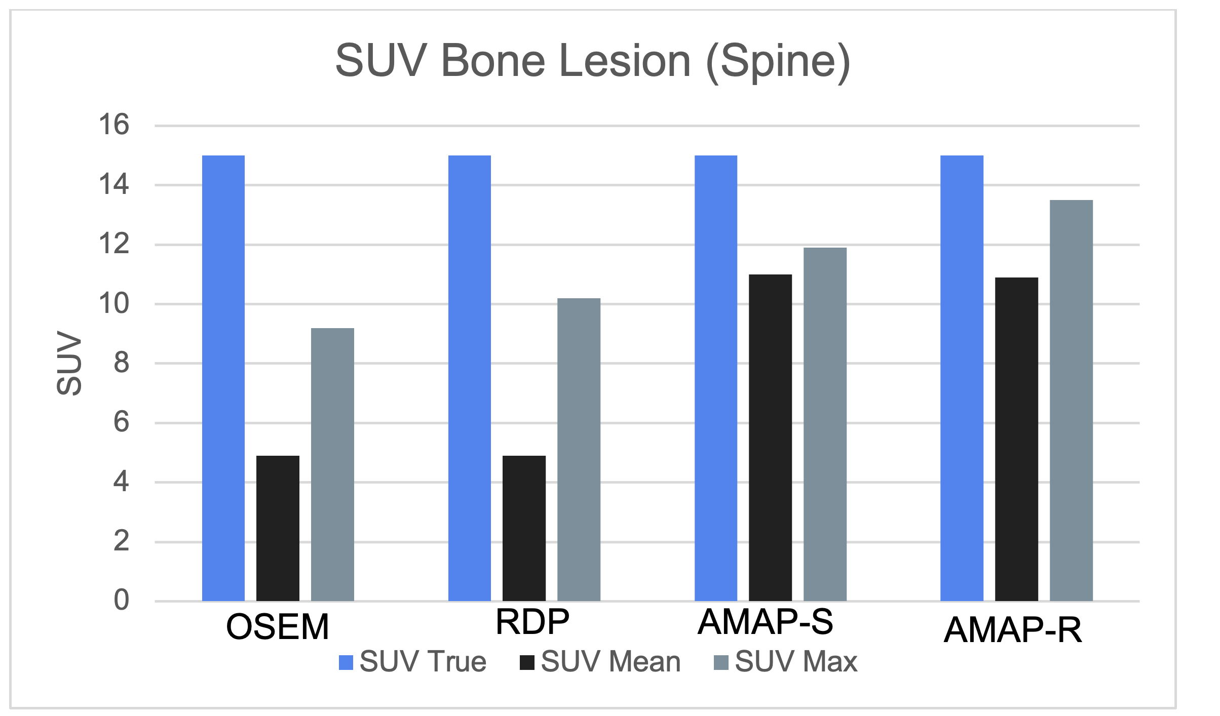
The authors concluded2, “The Bayesian methods with anatomical prior, especially the relative difference prior-based method (AMAP-R), outperformed OSEM and reconstruction without anatomical prior in terms of quantitative accuracy.”
1. Vuohijoki et al. Nuclear Medicine Communications (2023)
2. Kangasmaa et al. EJNMMI Physics (2021) 8:2)