Lung Lobe Quantification and Lung SPECT VQ
Anatomically accurate lung function and volumes with Lung Lobe Quantification and fast and easy Lung SPECT VQ image display and analysis.

Hermia applications for Pneumology
1. Lung Lobe Quantification
Improve outcomes for patients undergoing lung volume reduction surgery with accurate Lung Lobe Quantification method.
2. Lung SPECT VQ
Navigate, co-register, and analyse ventilation and perfusion data quickly and with ease.
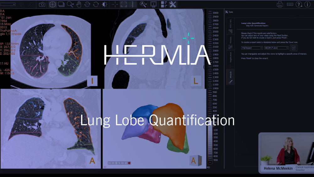
Lung Lobe Quantification
Lung Lobe Quantification™ achieves quick and precise prediction of function in each lobe by combining semi-automated CT segmentation of lung, airway, and lobar anatomy with functional image data.
PREOPERATIVE EVALUATION OF REGIONAL LUNG FUNCTION
Lung Lobe Quantification™ software provides an automated workflow for accurate and reliable surgical planning; preoperative evaluation of regional lung function is crucial to planning and predicting the outcomes of surgery.1 Individual identification of the most affected lobe in pulmonary emphysema before volume reduction procedures is essential to optimize the respiratory pump function,2 and prevents mistargeting a lobe that makes significant contribution to lung function.3
FAST AND RELIABLE QUANTITATIVE RESULTS
Processing takes 5-10 minutes per patient for a complete evaluation, compared with 45 minutes for manual segmentation and calculation. Excellent correlation was found with the results of manual segmentation methods and phantom studies in published validation work.4-5 A manually guided segmentation workflow is available for the most challenging cases.
EXTERNAL CT FOR ANATOMICAL DEFINITION
There is excellent concordance between lung perfusion results for hybrid SPECT/CT vs the non-hybrid approach.6
Use Lung Lobe Quantification™ to combine an existing diagnostic CT with conventional SPECT data to provide reliable lobar perfusion results without the need for additional radiation exposure to the patient.
3. Trahair E, Nowosinska E, Sizer N, Burniston M, Balan A, Lau K, O'Shaughnessy T, Sotiropoulos G, Waller D. Does 99mTc-MAA SPECT/CT have a role in evaluation of lung lobar perfusion in patients with chronic obstructive airway disease (COPD) prior to lung volume reduction surgery (LVRS). Nuclear Medicine Communications (2019) 40:393-453
5. Thillainathan A, Gregg S, Elsner A, Avondo J, Bailey J, Reyes E, Underwood S, Wechalekar K. Comparison of 2D planar and 3D SPECT-CT quantification of lung function in patients with lung cancer. Eur J Nucl Med Mal Imaging (2013) 40 (Suppl 2): Sl-S477 pp: 204-205
Lung Lobe Quantification
“Excellent concordance was found for 3D-quantification of relative lung perfusion when comparing a hybrid vs. non-hybrid approach.”
- Knollmann et al. Is hybrid SPECT/CT necessary for preinterventional 3D quantification of relative lobar lung function? European Journal of Hybrid Imaging (2018) 2:18
“Individual identification of the most affected lobe in pulmonary emphysema before volume reduction procedures is essential to optimize the respiratory pump function.”
- Schaefer WM et al.: Volume/perfusion ratio in pulmonary emphysema, Nuklearmedizin 1/2018
Fast and easy Lung SPECT VQ image display and analysis
This dedicated Lung SPECT VQ application allows you to co-register, navigate and analyse ventilation and perfusion data quickly and with ease:
- View and synchronously navigate tomographic ventilation and perfusion images
- Lung View provides a flexible display of transaxial, coronal, sagittal and MIP views of ventilation and perfusion, with synchronised triangulation and navigation. Automatically created subtraction and ratio images are displayed in TCS and MIP views, maintaining triangulation with ventilation and perfusion SPECT images
- Splash View provides a flexible slice-by-slice display of transaxial, coronal or sagittal views of ventilation and perfusion with synchronised triangulation and navigation
Hermia Lung VQ
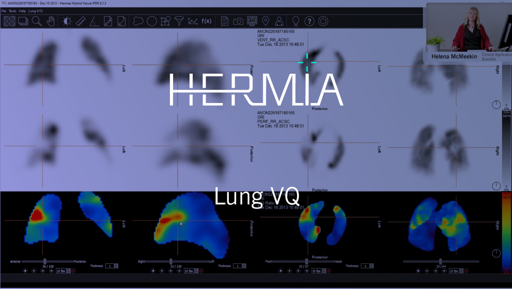
V/Q RATIO IMAGES TO HIGHLIGHT PERFUSION DEFECTS
Automatically create, display and synchronously navigate parametric normalised V/Q ratio images together with ventilation and ventilation-subtracted perfusion data. V/Q ratio (or 'quotient') images highlight areas of mismatch between ventilation and perfusion, indicating possible pulmonary embolism.
SUBTRACTION IMAGES TO CORRECT FOR OVER-VENTILATION
Ventilation counts are automatically subtracted from perfusion SPECT for clearer visualisation. Ventilation counts are decay corrected to account for the difference between acquisition times and then subtracted from the perfusion image when both are acquired with 99mTc. Subtraction is not performed when 81mKr ventilation images are loaded.
VQ SPECT/CT DISPLAY FOR INCREASED REPORTING CONFIDENCE
Combine information from multiple modalities to increase reporting confidence: fuse lung SPECT with local or external chest CT to correlate functional defects with anatomical information. Use the CT from a simultaneous SPECT/CT acquisition or import an external CT and automatically align it with the SPECT data.
Technical and Clinical Advantages of VQ SPECT Imaging
Dr. Charlotte Fowler at Guy St. Thomas Hospital in London gives a review of the technical and clinical advantages of VQ SPECT imaging.

Request more info or a demonstration
We would be happy to show you the many possibilities offered by HERMIA through a demonstration or to answer any questions you might have.
To support all your clinical workflows
Browse to read more about all the many possibilities offered by Hermia– our ALL-IN-ONE state-of-the-art software suite. You can pick and choose the specialities and tools adapted to your current clinical needs and scale up whenever new possibilities arise.
-
Read more
Nuclear Medicine Processing
Hermia offers the most comprehensive Nuclear Medicine processing toolkit on the market. Dedicated CE-marked, fully validated applications, for all clinical specialties and all associated image data types.
-
Read more
SPECT reconstruction
Hermia's SPECT reconstruction is optimized for speed and a wide range of procedures, radio-pharmaceuticals and collimators, making it possible to improve image quality while reducing dose and acquisition time from all your SPECT/CT cameras.
-
Read more
Neurology
Hermia Neurology has a fully automated 'single click' workflow. Data is processed, quantified and displayed within seconds with minimal user intervention.
-
Read more
Cardiology
State-of-the-art product line for cardiology, including third-party software with Invia Corridor4DM and Cedars-Sinai Cardiac Suite.