Anatomically guided reconstruction improves lesion quantitation and detectability in bone SPECT/CT
Vuohijoki, H, Constable C, Sahlberg AO.
Abstract
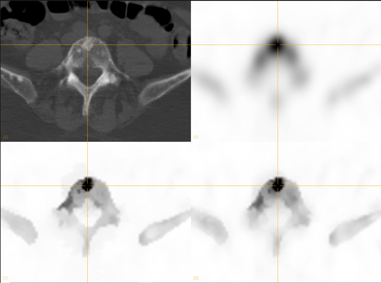
Bone single-photon emission computed tomography (SPECT)/computed tomography (CT) imaging suffers from poor spatial resolution, but the image quality can be improved during SPECT reconstruction by using anatomical information derived from CT imaging. The purpose of this work was to compare two different anatomically guided SPECT reconstruction methods to ordered subsets expectation maximization (OSEM) which is the most commonly used reconstruction method in nuclear medicine. The comparison was done in terms of lesion quantitation and lesion detectability. Anatomically guided Bayesian reconstruction (AMAP) and kernelized ordered subset expectation maximization (KEM) algorithms were implemented and compared against OSEM. Artificial lesions with a wide range of lesion-to-background contrasts were added to normal bone SPECT/CT studies. The quantitative accuracy was assessed by the error in lesion standardized uptake values and lesion detectability by the area under the receiver operating characteristic curve generated by a non-prewhitening matched filter. AMAP and KEM provided significantly better quantitative accuracy than OSEM at all contrast levels. Accuracy was the highest when SPECT lesions were matched to a lesion on CT. Correspondingly, AMAP and KEM also had significantly better lesion detectability than OSEM at all contrast levels and reconstructions with matching CT lesions performed the best. Quantitative differences between AMAP and KEM algorithms were minor. Visually AMAP and KEM images looked similar. Anatomically guided reconstruction improves lesion quantitation and detectability markedly compared to OSEM. Differences between AMAP and KEM algorithms were small and thus probably clinically insignificant.