The most comprensive toolkit on the market

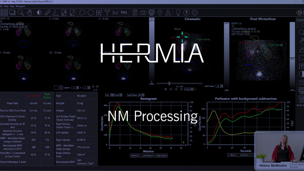
Nuclear Medicine Processing
HERMIA offers the most comprehensive Nuclear Medicine processing toolkit on the market. Dedicated and proven applications, for all clinical specialties and all associated image data types, that follow the same intuitive steps to facilitate easy adoption within your clinical workflow for:
Hermia Nuclear Medicine Processing
Comprehensive, end-to-end workflow for bone related indications
- Wholebody and localized planar and SPECT/CT image review
-
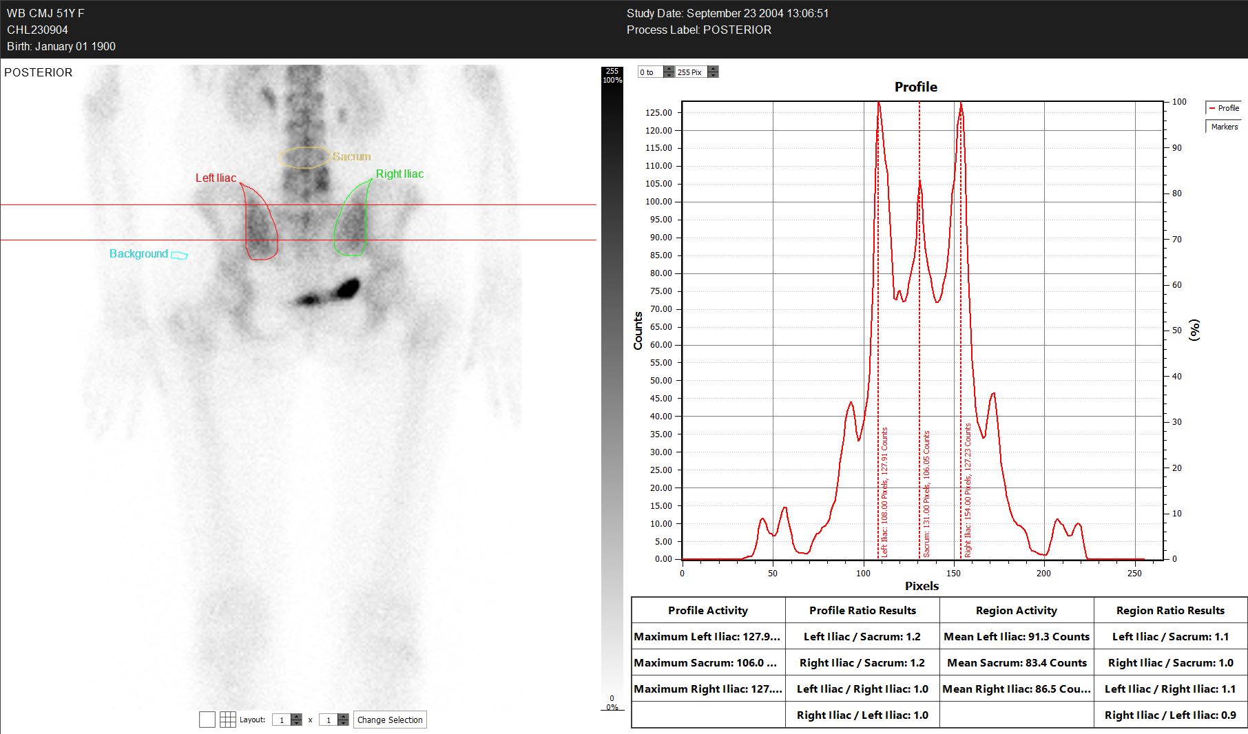
Dedicated Sacro-iliac Joint application.
- 3-Phase Bone Analysis
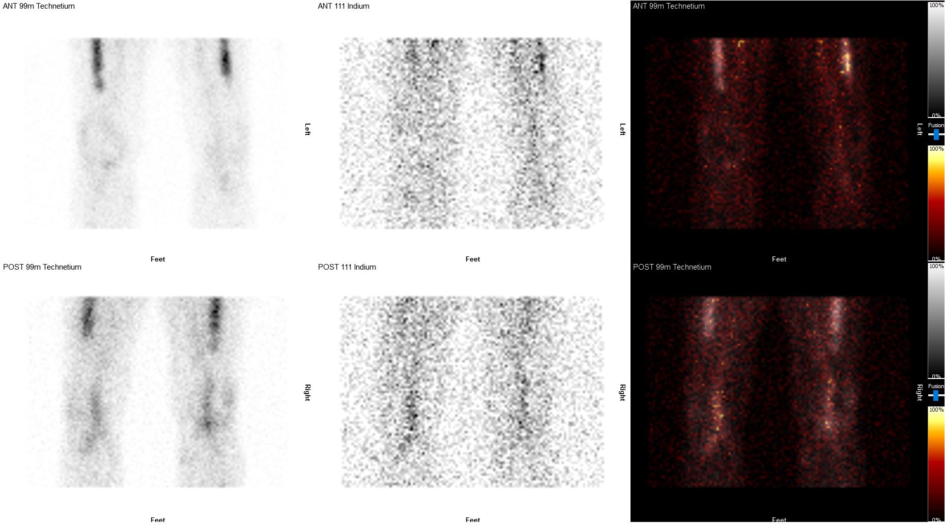
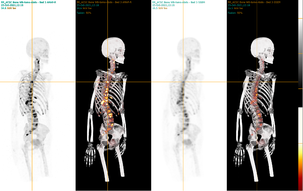
OSTEOLOGY
Comprehensive, end-to-end workflow of multi-bed reconstruction, image review and quantification in patients undergoing molecular imaging for bone related indications, including infection imaging, assessment of bone metastasis and sacroiliac joint pain.
Wholebody, localised planar and SPECT/CT image review is facilitated by user-friendly fully customisable viewers, supported by a wide range of tools for image manipulation and quantification at your fingertips.
The HERMIA bone indication CE marked applications include a dedicated Sacro-Iliac Joint application, facilitating fast semi-automated regional quantification with user-friendly retrospective region reload and edit functionality, along with a 3-Phase Bone Analysis application which supports efficient quantification of dynamic flow imaging alongside early and delayed planar timepoints.
Review and quantification for both single-visit and serial bone planar wholebody, SPECT/CT and PET/CT within HERMIA is fast and intuitive, supporting efficient, reproducible reporting workflows and increasing clinical confidence and clarity.
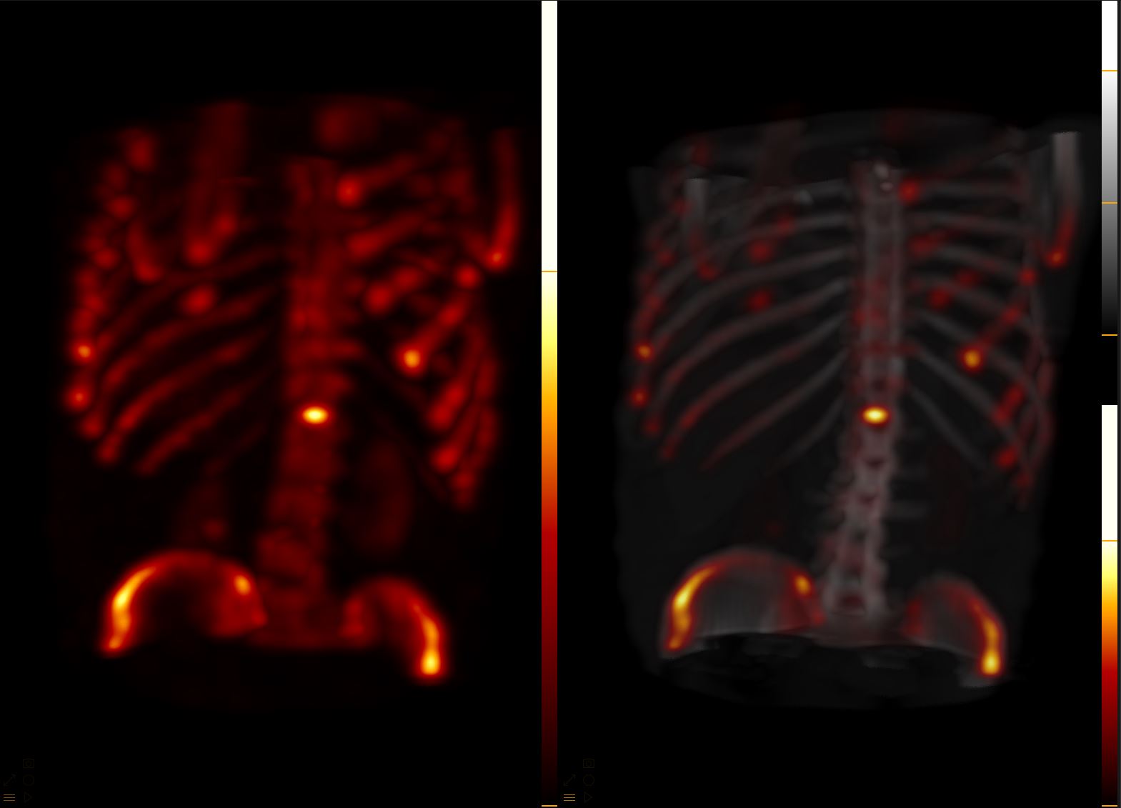
With HERMIA advanced reconstruction techniques, SUV-SPECT bone quantification can become part of your routine patient workflow!
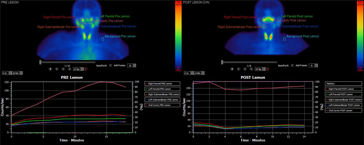
Intestine functionality, saliva production
- Gastric emptying (Dynamic and planar)
- Oesophageal transit and reflux
- Colonic Transit
- Salivary Gland Analysis
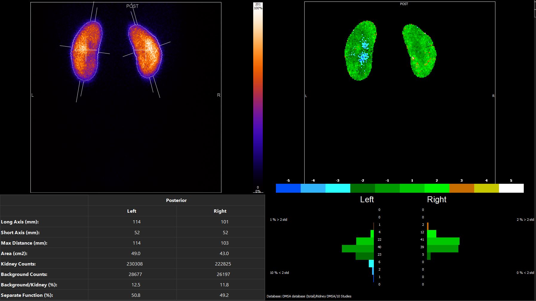
A comprehensive toolkit for the kidney functionality
- DMSA
- Renogram
- Duplex Kidney Analysis
- Renal Patlak Analysis
- Advanced Renal Analysis
NEPHROLOGY
The comprehensive Nephrology toolkit within Hermia supports DMSA SPECT reconstruction and advanced quantification for all your Nuclear Medicine nephrological investigational needs, tailored to your custom acquisition protocols and personalised to your reporting requirements.
Hermia offers the flexibility to process all patient cohorts, exhibiting all urological anatomical variations including duplex kidneys and transplant organs, within one user-friendly adaptable platform.
The Hermia DMSA toolkit facilitates accurate determination of split function for planar and SPECT DMSA studies and supports geometric mean quantification. Enhanced tools include automated regional delineation and access to a normal database for statistical deviation comparison in paediatric patients.
Hermia Renogram supports advanced processing techniques with extensive quantification options including Patlak background correction, Transit time analysis, NORA and Output Efficiency. Geometric mean and post-micturition/delayed analysis are supported, automatically representing data according to relative acquisition times and effortlessly accommodating any deviations from standard protocol that may result in interrupted acquisitions and challenging reporting scenarios.
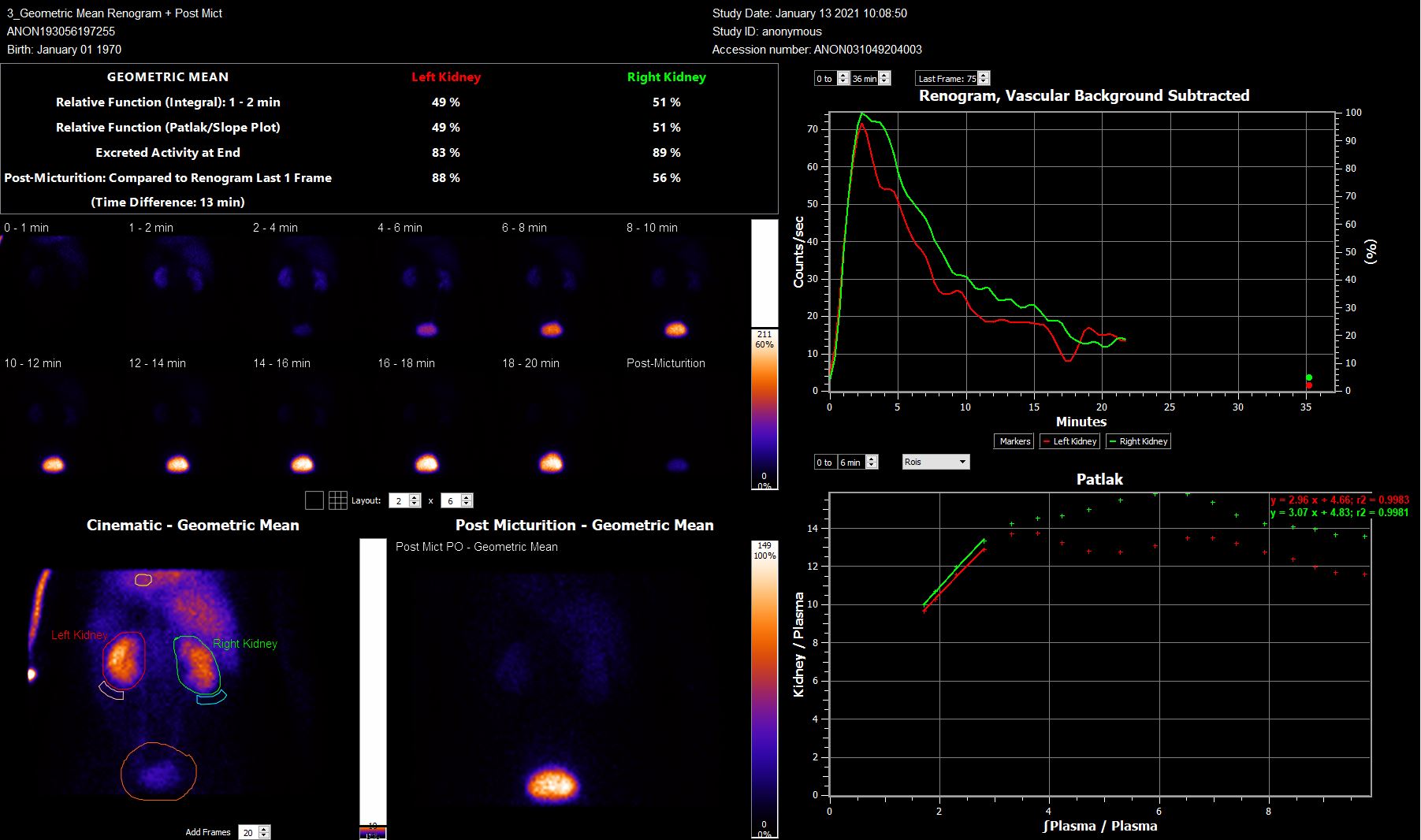
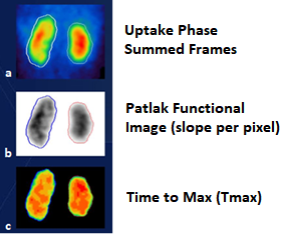
Hermia seamlessly integrates with Renal Curve™ offering extended renography and post micturition analysis. Improved deconvolution algorithms within Renal Curve™ result in clearer plateaus on retention function graphs and reconvolution curves support quality control of deconvolution analysis. Signal to noise time activity curve smoothing and pseudo-planar functional image generation, from summed background corrected frames during the kidney uptake phase, Patlak slope and Time to Max, allows assessment on a per pixel basis for comprehensive functional analysis.
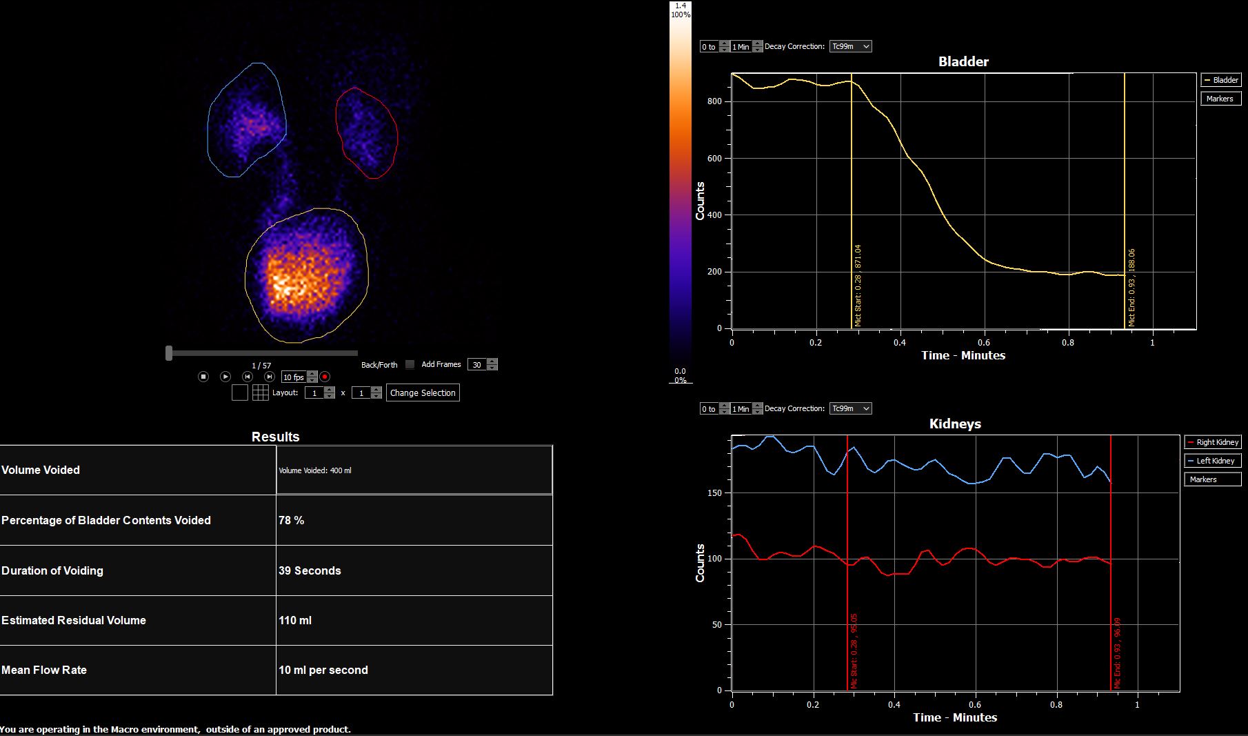
Radionuclide Cystography quantitative analysis within Hermia is achieved with significant flexibility, including options for custom macro creation to expand the analysis to best match your workflows and protocol.
Liver function, Prep for surgery, Gallbladder functionality
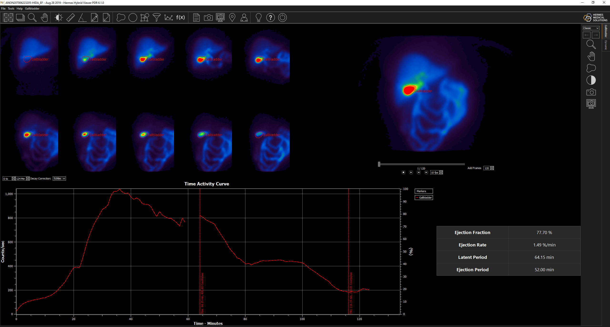
- HIDA
- Gallbladder ejection fraction
- Future Remnant Liver Function (FRLF) analysis
“Standardization of the dynamic Tc-99m Mebrofenin hepatobiliary scintigraphy (HBS) combined with SPECT/CT technique and the use of a shared imaging software as Hermia NM Processing with remote access seems in our opinion of utmost importance to enable comparisons among different centers.”
- Matteo Serenari MD, Chiara Bonatti MD, Malpighi Hospital, Alma Mater Studiorum, University of Bologna – Cinzia Pettinato MD, Orsola-Malpighi Hospital, Bologna, Italy
All the tools needed for quantification of the thyroid function and for the localization of the parathyroid adenoma.
- Assessment of thyroid uptake
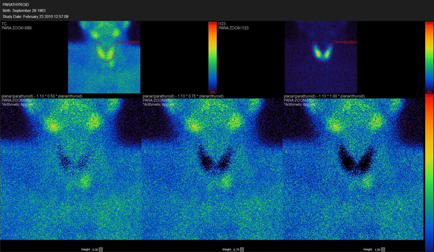
- Parathyroid Substraction (Planar and SPECT)
To support all your clinical workflows
Browse to read more about all the many possibilities offered by Hermia– our ALL-IN-ONE state-of-the-art software suite. You can pick and choose the specialities and tools adapted to your current clinical needs and scale up whenever new possibilities arise.
-
Read more
Pneumology
Anatomically accurate lung function and volumes with Lung Lobe Quantification and fast and easy Lung SPECT VQ image display and analysis
-
Read more
Cardiology
State-of-the-art product line for cardiology, including third-party software with Invia Corridor4DM and Cedars-Sinai Cardiac Suite.
-
Read more
Neurology
Hermia Neurology has a fully automated 'single click' workflow. Data is processed, quantified and displayed within seconds with minimal user intervention.
-
Read more
Future Remnant Liver Function
Hermia Software Analysis allows you to obtain quantitative results to assess post-surgical future remnant liver function in minutes.